October 10, 2016
Scanning EM of cryptococci
Wild type cryptococci imaged by scanning EM. Image by Felipe Santiago-Tirade. See Santiago-Tirado et al, 2016....
Read More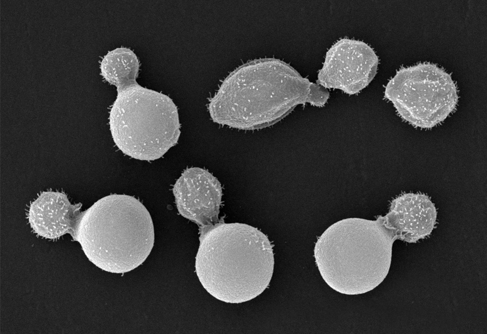
Wild type cryptococci imaged by scanning EM. Image by Felipe Santiago-Tirade. See Santiago-Tirado et al, 2016....
Read More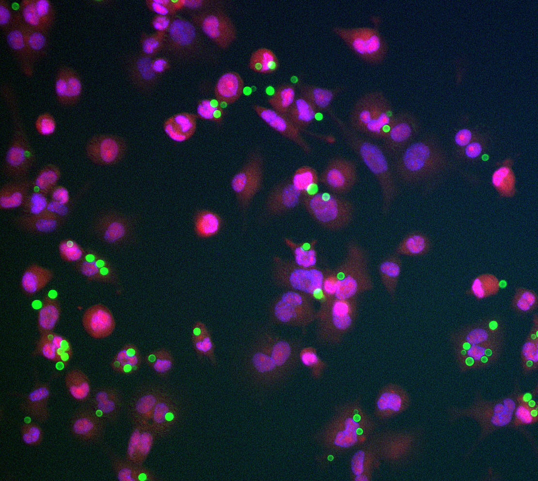
Cryptococcal cells (green) interacting with the human THP-1 phagocytes (nuclei, pink; cytosol, red). Image by Deepa Srikanta. See Srikanta et al, 2011....
Read More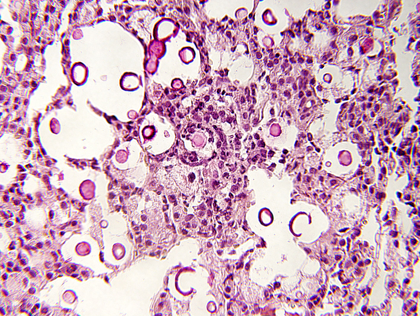
Mucicarmine stain of a lung section from a mouse infected with C. neoformans, showing host inflammation, tissue destruction, and abundant fungi (walls stained maroon). See Gish et al, 2016....
Read More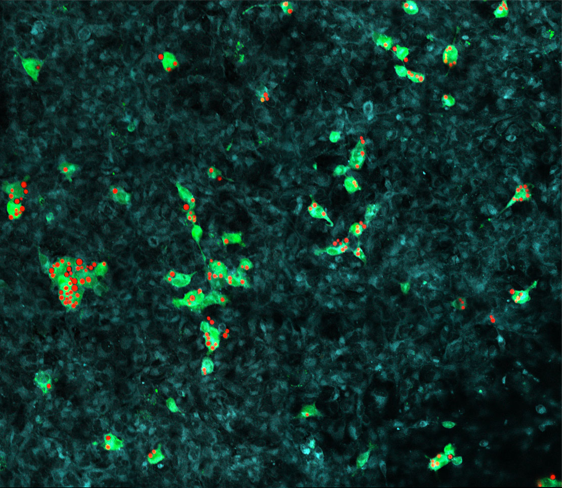
Human phagocytes (green) with engulfed cryptococci (red) interacting with a layer of brain endothelial cells (blue). Image by Felipe Santiago-Tirado. See Santiago-Tirado et al, 2017....
Read More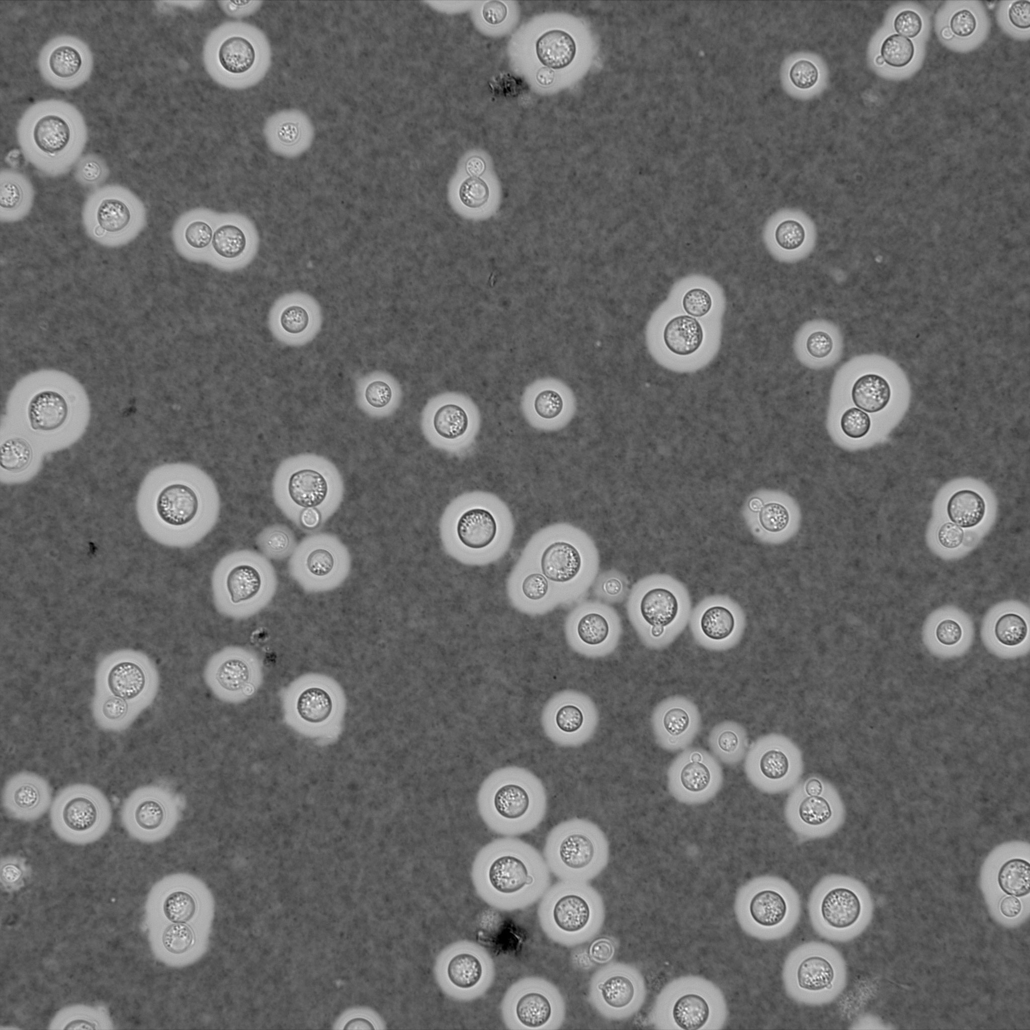
Light micrograph of cryptococci induced to form capsule and then mixed with India ink. The space occupied by the capsule shows as a clear space between the gray background of the ink particles and the refractile edge of the cell....
Read More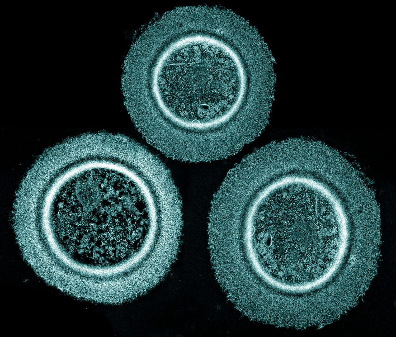
Thin section electron micrograph of wild type cryptococci after growth in capsule-inducing medium. Image by Aki Yoneda, colored by Wandy Beatty....
Read More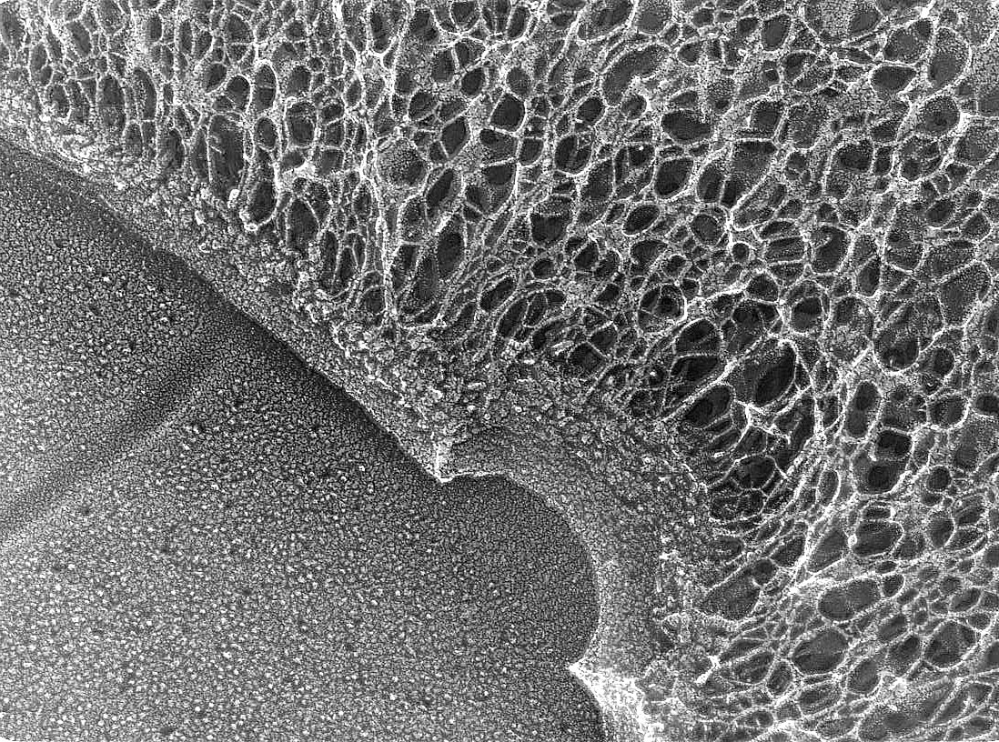
Quick-freeze deep-etch image of the edge of a budding cryptococcal cell, showing a region of the plasma membrane (bottom left) bounded by the bilayered cell wall, with capsule fibers extending to the right from both the parent cell and a newly forming bud. Image by Tamara Doering and John Heuser....
Read More
Thin section of a budding C. neoformans that accumulates secretory vesicles because it has a sav1 mutation. Image by Aki Yoneda. See Yoneda et al, 2006....
Read More