Search
Infected lung tissue – Doering Lab
7594
portfolio-item-template-default,single,single-portfolio-item,postid-7594,eltd-core-1.0,woly child-child-ver-1.0.0,woly-ver-1.0,eltd-smooth-scroll,eltd-smooth-page-transitions,eltd-mimic-ajax,eltd-grid-1200,eltd-blog-installed,eltd-follow-portfolio-info,eltd-main-style1,eltd-disable-fullscreen-menu-opener,eltd-header-standard,eltd-sticky-header-on-scroll-down-up,eltd-default-mobile-header,eltd-sticky-up-mobile-header,eltd-menu-item-first-level-bg-color,eltd-dropdown-slide-from-top,eltd-,eltd-header-standard-disable-transparency,eltd-fullscreen-search eltd-search-fade,wpb-js-composer js-comp-ver-4.12,vc_responsive

Infected lung tissue

Infected lung tissue

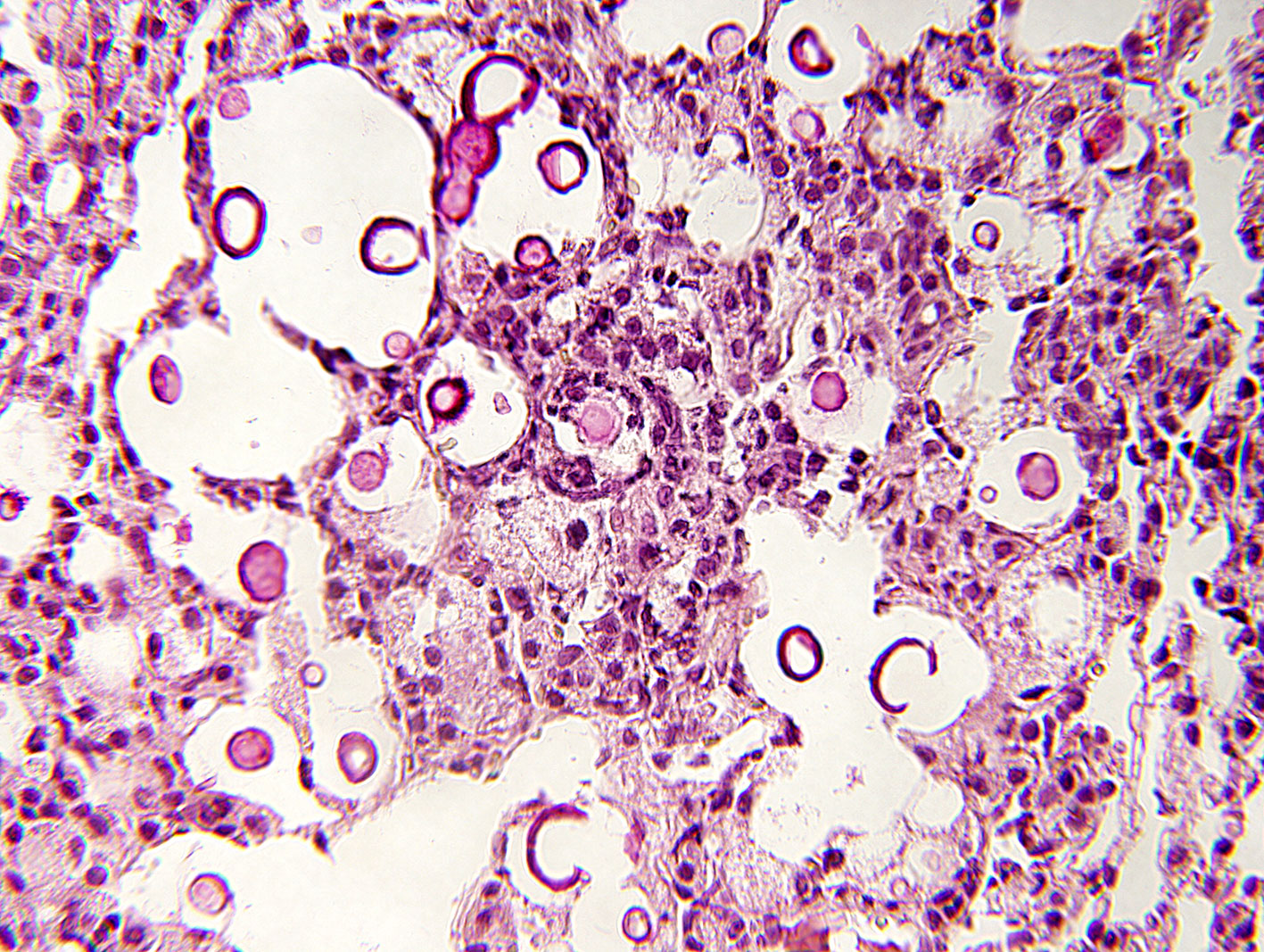
Mucicarmine stain of a lung section from a mouse infected with C. neoformans, showing host inflammation, tissue destruction, and abundant fungi (walls stained maroon). See Gish et al, 2016.

Follow Us!